Tools
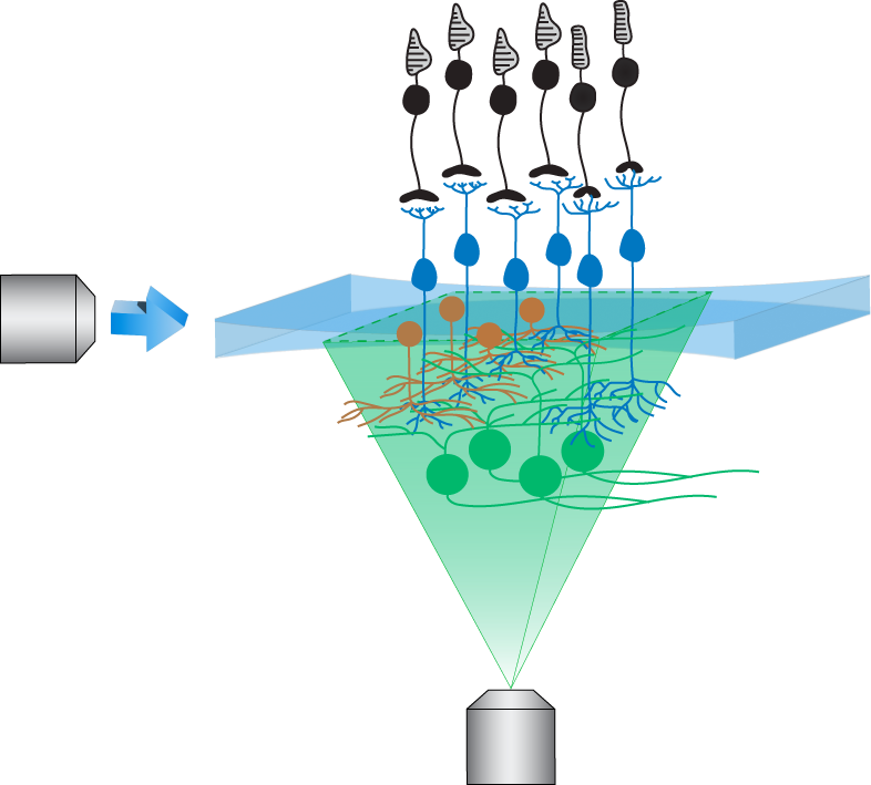
Light sheet imaging of ex-vivo retina

Built on the foundation of restricted planar imaging, light-sheet fluorescence imaging allows measurements of neural activity at single-cell resolution across large swathes of ex-vivo retina (read more). We are refining these tools to study precise signal communication within neuronal compartments such as dendrites and axon terminals, and across synaptic layers, in genetically targeted interneurons and retinal ganglion cells.
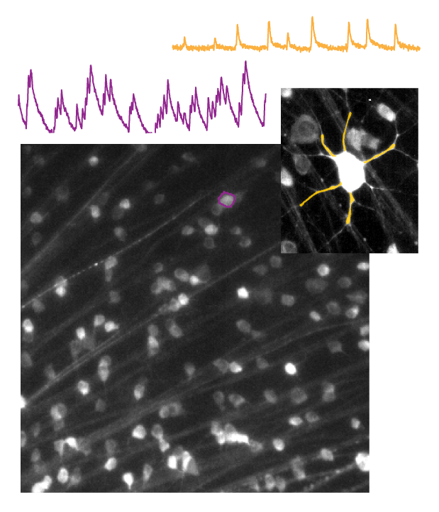
Image analysis

Image analysis is an essential component in studies assessing functional properties of retinal neurons to studies attempting early detection of retinal diseases. We are developing and deploying new methods of image analysis into our studies, to reliably extract neural activity from fluorescence dynamics across large populations of retinal neurons and interpreting patterns of activity in the context of visual stimuli. Check out our 1-photon image analysis pipeline.
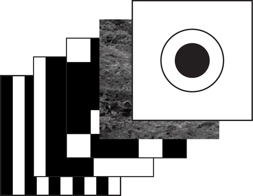
Visual stimuli

We are working on expanding the repertoire of visual stimuli from artificial to natural images for use in experiments. These stimuli will allow us to tease apart specialized functions that retinal circuits implement.